Emergency Ultrasound
Format
This pre-course is offered in room 143, at Palais des Congrès de Paris, France on Thursday 24 September, 08:00 -18:00 CEST.
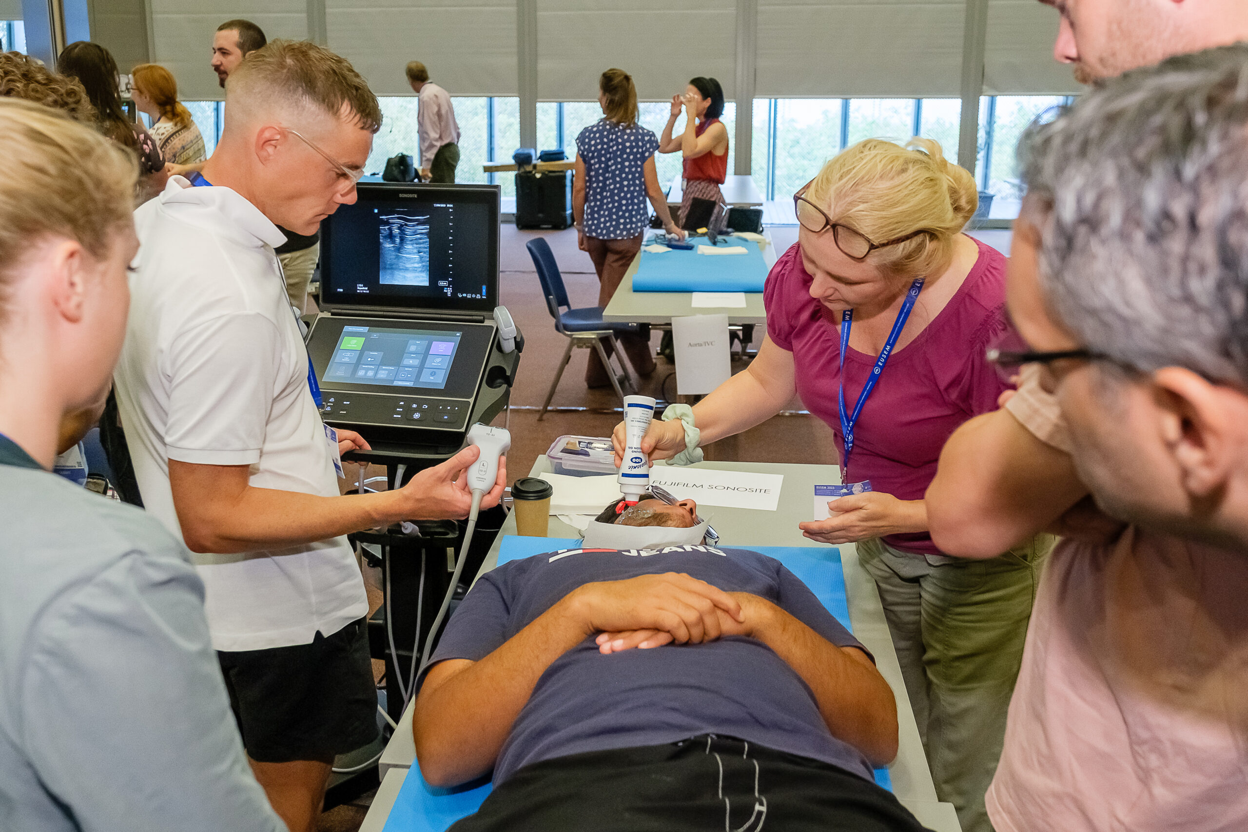
This Point-of-Care Ultrasound (POCUS) training offers a dynamic learning experience for emergency physicians, combining foundational knowledge, hands-on practice, and expert-led instruction. By choosing relevant modules for their individual clinical practice participants will enhance their diagnostic and procedural ultrasound skills, gaining confidence in applying POCUS to improve patient outcomes in emergency care settings. The course will be submitted for CME accreditation by the EACCME®.
For who
Emergency physicians (consultants, trainees, emergency medicine physician assistants), paramedics and nurses.
Module 1 – Ultrasound guided nerve blocks in trauma
This module will provide an introduction to ultrasound guided nerve and fascial plane blocks deemed of high value to emergency physicians in their daily practice. By
providing a foundation to work on, the participant will then be able to apply the principles and skills to any other blocks that are useful in their scope of practice.
Learning Objectives
- Foundations of regional anaesthesia
- know the nerve, see the nerve, see the needle, do the block.
- Practice needling technique including some challenges to test your skills!
- Blocks covered in the module with practical sessions.
o Fascia Iliac compartment blocks for hip fractures–suprainguinal and
infrainguinal.
o Femoral nerve.
o Popliteal.
o Chest wall blocks.
§ Serratus Anterior Plane.
§ Erector Spinae Plane. - Overview of upper limb regional anaesthesiae options
Module 2 – POCUS in musculoskeletal emergencies
This course provides hands-on training in musculoskeletal (MSK) point-of-care ultrasound (POCUS) for joint, tendon, muscle, and soft tissue evaluation relevant for emergency physicians. Participants will learn to identify pathologies, perform ultrasound-guided procedures, and enhance their diagnostic skills.
Learning Objectives
POCUS of the Joints:
- Identify joint eQusion in the ankle, knee, hip, shoulder, and elbow using POCUS.
- Perform ultrasound-guided joint aspiration and infiltration.
- Utilize POCUS for evaluating shoulder dislocations.
POCUS of the Tendons and Muscles:
- Diagnose ruptures of the Achilles tendon, patellar tendon, quadriceps tendon,
and biceps tendon. - Identify rupture of the medial head of M. gastrocnemius (Tennis Leg).
POCUS of the Soft Tissue:
- DiQerentiate soft tissue swelling using POCUS.
- Perform ultrasound-guided foreign body removal.
- Conduct a tibialis posterior nerve block for foreign body removal from the sole of
the foot.
Module 3 – Basic focused cardiac POCUS for emergency physicians
In this module, designed specifically for emergency physicians, we explore the
theoretical and practical aspects of cardiac ultrasound. Participants will learn basic
anatomy and views, work on hand-eye coordination, interpret ultrasound findings
comparing normal and pathological images, and integrate results into clinical reasoning
in time-sensitive pathologies.
Learning Objectives
- getting familiar with basic cardiac views and describing the relevant anatomy of
- every view: subcostal, parasternal long and short axis, apical four, five and two
chamber view - name diQerent regions of the left ventricular muscle and describe diQerent basic
ways to assess the contractility - assess left ventricular contractility in diQerent views by eyeballing
- compare and measure chamber size in A4C view
- assess the size of the right ventricle and motion of the septum in diQerent views
- IVC measurement pitfalls and interpretation
- pericardial eQusion and tamponade
Module 4 – Advanced focused cardiac POCUS for emergency physicians
Participants will delve into advanced techniques, mastering, including comprehensive, semi-quantitative and even quantitative assessment of cardiac function, identification of structural abnormalities, and evaluation of hemodynamic status. By the end of the course, participants will be taught on tips for integrating all of these applications into their daily clinical practice for critical decision-making in urgent scenarios.
Learning Objectives
- Assess left ventricular systolic function using multiple echocardiographic views, including visual estimation, MAPSE, EPSS, and quantitative methods such as Teichholz and Simpson.
- Evaluate the mitral valve in B-mode and with colour Doppler to identify severe mitral regurgitation.
- Assess right ventricular systolic function using chamber size, TAPSE, and septal motion, and distinguish features suggestive of chronic right ventricular strain.
- Measure tricuspid regurgitation jet velocity (TRV) and pulmonary acceleration time (PAT) in the assessment of suspected pulmonary embolism.
- Measure LVOT VTI to calculate cardiac output and assess haemodynamic changes during a passive leg raise test.
- Learn the principles and technique of echocardiography-guided pericardiocentesis.
Faculty
Course Director: Dr. Senad Tabakovic, (CH)
Co-Directors: Dr. Eftychia Polyzogopoulou (GRC) and Paul Van Overbeeke (NL)
- Tomas Villen
- Sánchez Sendín Domingo
- Lorang Felix
- Majeed Azam
- Keranovic Adis
- Nassralah Nagib
- Ivan Gornik
- Berdouk Sabrina
- Abdolghader Paknyat
- Cardenas Linder
- Grinna Mats
- Aslaner Mehmet Ali
- Haesendonck Ruben
- Chris Yap
- Chin Eric
- Osterwalder Joseph
- Radulovic Bojana
- Croft Peter
- Matthies Ashley
- Cloete David
- Hoffmann Beatrice
- Bajic Slaven
- Atesli Zeki
- Sukul Prem
Registration
Each participant may select either one module for half a day or two modules for full day.
The price for 1/2 day is € 275 (including 10% VAT)
The price for full day is € 475 (including 10% VAT)
Please register through your MyEUSEM account by clicking on the EUSEM Congress registration button. From the Pre-courses tab you can select the pre-courses you would like to join.
Programme
Module 1 and module 3 are running in parallel. Module 2 and module 4 are running in parallel.
- Thursday 24 September – Module 1
- Thursday 24 September – Module 2
- Thursday 24 September – Module 3
- Thursday 24 September – Module 4
Module 1 – Ultrasound guided nerve blocks in trauma
| Time | 24 September |
| 08:15-08:30 | IIntro to the day and Needling top tips |
| 08:30-09:30 | Needling challenges (hands on) |
| 09:30-09:45 | Blocks for lower limb fractures |
| 09:45-10:45 | Hands on |
| 10:45-11:00 | Coffee Break |
| 11:00-11:15 | Chest wall injuries blocks |
| 11:15-12:15 | Hands on |
| 12:15-12:30 | Upper Limb RA options and Close |
Module 2 – Ultrasound in musculoskelettal emergencies
| Time | 24 September |
| 13:30-13:35 | Welcome and Introduction |
| 13:35-13:50 | Knee Effusion/Aspiration, Patellar Tendon Rupture, Quadriceps Tendon Rupture; Hip effusion/aspirationn |
| 13:50-14:40 | Scanning |
| 14:40-14:55 | Ankle and Lower Leg (Achilles Tendon Rupture, Ankle Effusion/Aspiration, Syndesmosis Rupture, Distal Malleolar Fractures, Medial Gastrocnemius Rupture) |
| 14:55-15:45 | Scanning |
| 15:45-16:00 | Coffee break |
| 16:00-16:15 | Shoulder and Elbow (Effusion/Aspiration, Shoulder Dislocation, Biceps Tendon Rupture, Supraspinatus Tendon and Subacromial Bursa Infiltration) |
| 16:15-17:00 | Scanning |
| 17:00-17:15 | Soft Tissue Swelling and US-Guided Foreign Body Removal, Tibialis Posterior Block |
| 17:15-18:00 | Scanning and close |
Module 3 – Basic focused cardiac POCUS for emergency physicians
| Time | 24 September |
| 08:15-08:25 | Introduction |
| 08:25-08:40 | Basic views: SX, PLAX, PSAX, A4 and IVC |
| 08:40-09:40 | Hands on |
| 09:40-09:55 | LV basic assessment |
| 09:55-10:25 | RV basic assessment |
| 10:25-10:40 | Coffee break |
| 10:40-11:40 | Hands on |
| 11:40-11:55 | Pericardial effusion and tamponde |
| 11:55-12:40 | Hands on and close |
Module 4 – Advanced focused cardiac POCUS for emergency physicians
| Time | 24 September |
| 13:30-13:40 | Introduction |
| 13:40-13:55 | Doppler for emergency physicians: C, PW, Cw, TDI |
| 13:55-14:10 | Advanced LV systolic assessment |
| 14:10-14:25 | Advanced RV systolic assessment |
| 14:25-15:25 | Hands on |
| 15:25-15:40 | Coffee break |
| 15:40-15:55 | Basic hemodynamics: from VTI to CO |
| 15:55-16:55 | Hands on |
| 16:55-17:10 | Echo guided pericardiocentesis |
| 17:10-18:00 | Hands on Interactive cases and close |