
Emergency Ultrasound Pre-course
Format
This pre-course is offered in room Schubert 1, at VIECON, Vienna on Saturday 27 September, 08:00 -18:30 CET and Sunday 28 September, 08:00 – 12:30 CET
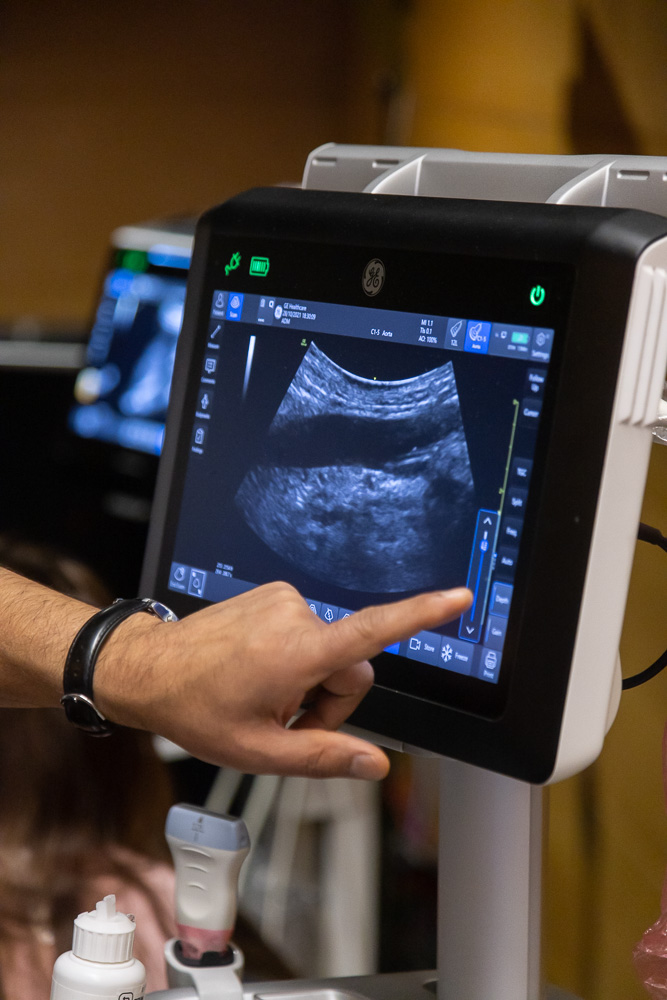
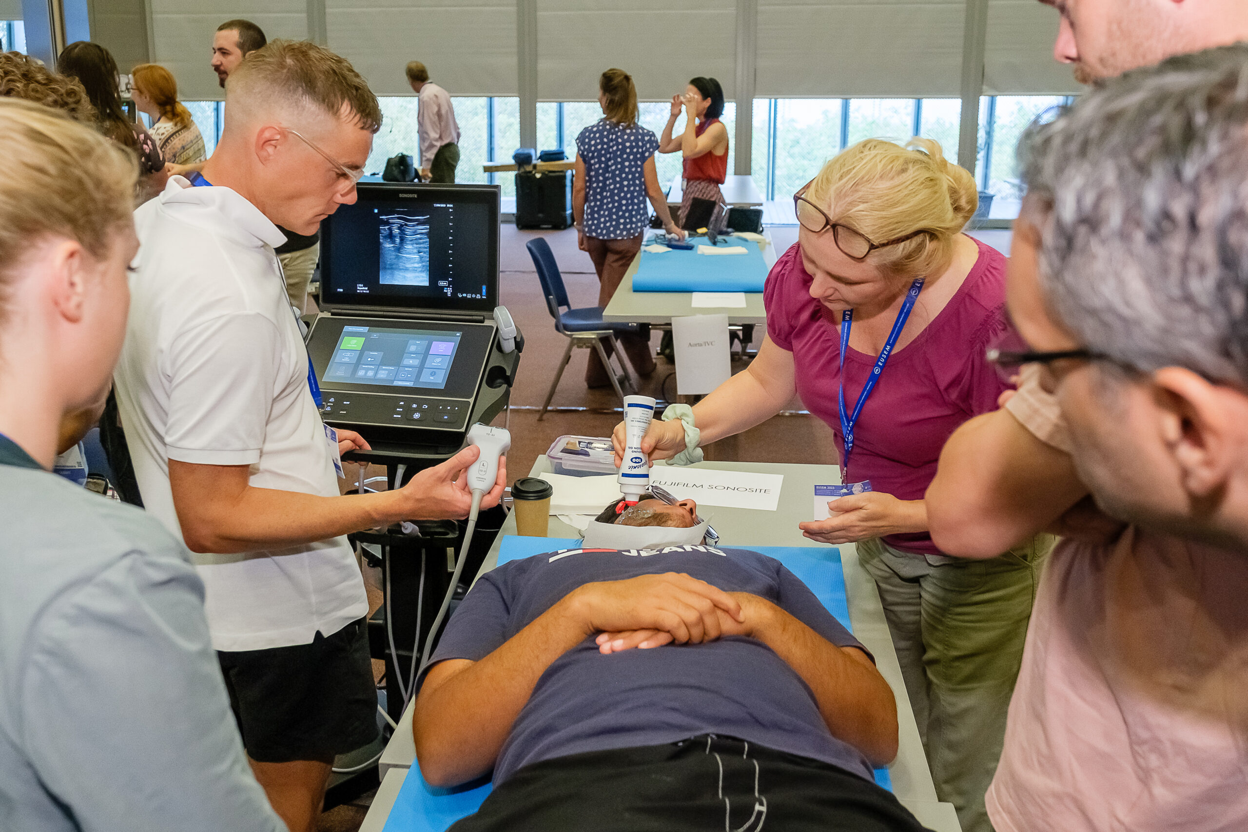
This Point-of-Care Ultrasound (POCUS) training offers a dynamic learning experience for emergency physicians, combining foundational knowledge, hands-on practice, and expert-led instruction. By choosing relevant modules for their individual clinical practice participants will enhance their diagnostic and procedural ultrasound skills, gaining confidence in applying POCUS to improve patient outcomes in emergency care settings. The course is accredited with 12 CMEs by the EACCME®.
For who
Emergency Physicians (consultants, trainees, Emergency Medicine Physician Assistants)
Saturday 27 September
Module 1 – POCUS in abdominal emergencies
This module provides hands-on training and theoretical knowledge in emergency ultrasound (POCUS) for the evaluation of abdominal and genitourinary emergencies. Participants will develop proficiency in identifying key ultrasound findings, performing targeted assessments, and conducting ultrasound-guided procedures for rapid diagnosis and patient management.
Learning Objectives
Cholecystitis/gallstones/biliary three
- How to find the gallbladder?
- How to assess for stones?
- How and where to assess the wall? What signs to look for in cholecystitis?
- How to find the DHC and where to measure the diameter?
Ileus
- How to perform the assessment of the small bowel and scan the stomach?
- How to assess the diameter and kinesis of the small bowel?
- What are US-signs of small bowel obstruction?
Free fluid in non-trauma patient and free air
- Where to look for free intraperitoneal fluid in a non-trauma patient?
- How to perform an US-guided paracentesis?
- Where to look for free air and which signs to look for?
Appendicitis and diverticulitis
- How to find the cecum and look for an inflamed appendix?
- How to find the sigma and colon descendens and what are the signs of an inflamed colon/ diverticula?
Aorta and retroperitoneum
- How to identify AAA with POCUS?
- How to look for a retroperitoneal hematoma with ultrasound?
Urolithiasis/pyelonephritis
- How to look for hydronephrosis?
- How to assess the narrowings of the ureter?
- Doppler settings for the Twinkling artifact
- How to measure stone diameter?
- What to look for in pyelonephritis?
- How to use Doppler to assess for kidney infarction?
Module 2 – focused cardiac POCUS for emergency physicians
In this module, designed specifically for emergency physicians, we explore the theoretical and practical aspects of cardiac ultrasound. In the morning participants will learn basic anatomy and views, work on hand-eye coordination, interpret ultrasound findings comparing normal and pathological images, and integrate results into clinical reasoning in time-sensitive pathologies. In the afternoon participants will delve into advanced techniques, mastering, including comprehensive, semi-quantitative and even
quantitative assessment of cardiac function, identification of structural abnormalities, and evaluation of hemodynamic status. By the end of the course, participants will be taught on tips for integrating all of these applications into their daily clinical practice for critical decision-making in urgent scenarios.
Learning Objectives
- getting familiar with basic cardiac views and describing the relevant anatomy of every view: subcostal, parasternal long and short axis, apical four, five and two chamber view
- name different regions of the left ventricular muscle and describe different basic ways to assess the contractility
- compare and measure chamber size in A4C view
- assess left ventricular contractility in different views by eyeballing
- assess the size of the right ventricle and motion of the septum in different views
- IVC measurement pitfalls and interpretation
- pericardial effusion and tamponade
- assess left ventricular contractility in different views by eyeballing, MAPSE, EPSS and Teichholz
- assess the mitral valve in B mode and with color for severe mitral regurgitation
- assess right ventricular contractility in different views by chamber size, TAPSE, septum motility and color over the tricuspide valve
- measure LVOT VTI and calculate CO and its changes during passive leg raise test
- learn how to perform an echo guided pericardiocentesis
Module 3 – PoCUS for lung emergencies and fractures of the chest
Lung and chest wall sonography is the newest discipline of point-of-care ultrasound for common and life-threatening emergencies. Its diagnostic accuracy far surpasses that of the stethoscope and chest x-ray. It is not only about to replace both, but also represents a valid alternative to computed tomography for several indications. Our workshop is intended for all those who do not want to miss out on this development. The only prerequisites are basic knowledge and familiarity with ultrasound knobology.
Learning Objectives
- physical background and the sonoanatomy:
- basic ultrasonic tools to examine accurately the lung and the chest wall. The focus is on the pleural line, horizontal reverberations and mirror artifacts, lung sliding, pulmonary pulse, costodiaphragmatic recess, ribs and sternum as well as most important pitfalls using these ultrasound applications
- examination technique, the settings in B-mode, M-mode and color
Doppler for the optimal visualization of the sonoanatomy and the avoidance
of pitfalls - interactive cases visualising pathologies, based on two forms of visualization: directly visualizable subpleural parenchymal changes and indirectly visualizable interstitial pathologies and aeriation of the lung by vertical artifacts with an update on the current state of knowledge, the nomenclature and ultrasonic presentation. In detail:
- pneumothorax
- pleural effusion
- pleuritis
- lobar pneumonia
- pulmonary embolism
- atelectasis
- rib and sternal fractures
- pulmonary edema
- pulmonary contusion
- pearls and pitfalls with the experts: the experts are going to discuss pearls and pitfalls of the different scanning techniques with the focus on the implementation of the scans in the clinical practice
Sunday 28 September
Module 4 – Module POCUS in musculoskeletal emergenices
This course provides hands-on training in musculoskeletal (MSK) point-of-care ultrasound (POCUS) for joint, tendon, muscle, and soft tissue evaluation relevant for emergency physicians. Participants will learn to identify pathologies, perform ultrasound-guided procedures, and enhance their diagnostic skills.
Learning Objectives
- POCUS of the Joints:
- Identify joint effusion in the ankle, knee, hip, shoulder, and elbow using POCUS.
- Perform ultrasound-guided joint aspiration and infiltration.
- Utilize POCUS for evaluating shoulder dislocations.
- POCUS of the Tendons and Muscles:
- Diagnose ruptures of the Achilles tendon, patellar tendon, quadriceps tendon, and biceps tendon.
- Identify rupture of the medial head of M. gastrocnemius (Tennis Leg).
- POCUS of the Soft Tissue:
- Differentiate soft tissue swelling using POCUS.
- Perform ultrasound-guided foreign body removal.
- Conduct a tibialis posterior nerve block for foreign body removal from the sole of the foot.
Module 5 – Ultrasound Guided Vascular Access
The objectives of this module are to provide participants with an up-to-date review of the latest developments in ultrasound-guided vascular access techniques, including peripheral lines, midlines, and central lines.
Learning Objectives
- Describe normal anatomy and anatomical variants relevant to ultrasound-guided vascular access, and differentiate between arteries and veins.
- Explain all steps of ultrasound guidance, including prescan, equipment selection, puncture technique, evaluation, and documentation.
- Demonstrate ultrasound needle coordination for successful vascular puncture.
- Describe ultrasound-guided puncture techniques using the longitudinal (inplane) and transverse (out-of-plane) approaches.
- Discuss special considerations for central venous line placement (internal jugular, femoral, and subclavian) and for peripheral and midline catheter placement (forearm, upper arm, basilic, and cephalic veins).
- Engage in hands-on training with a focused, interactive approach using Blue Phantoms, manikins (internal jugular, femoral, and upper limb), and live models.
Module 6 – Blocks in the ED
This module will provide an introduction to ultrasound guided nerve and fascial plane blocks deemed of high value to emergency physicians in their daily practice. By providing a foundation to work on, the participant will then be able to apply the principles and skills to any other blocks that are useful in their scope of practice.
Learning Objectives
- Foundations of regional anaesthesia
- Practice needling technique including some challenges to test your skills!
- Blocks covered in the module with practical sessions:
- Upper limb blocks (Median/Radial/Ulnar)
- Lower limb blocks (Fascia Iliac compartment blocks for hip fractures – suprainguinal and infrainguinal/Femoral nerve blocks for femoral fractures/Popliteal & Adductor Canal blocks for tibial and fibular fractures)
- Chest wall blocks (Serratus Anterior Plane/Erector Spinae Plane)
Faculty
Course Director: Dr. Senad Tabakovic, (CH)
Co-Directors: Dr. Paul Overbeeke, (NL) Dr. Effie Pol, (GRC)
Module leads:
Focussed heart: Tomas Villen (SPA) Focussed lung: Joseph Osterwalder (SUI) and Alexander Spiel (AUT) Focussed abdomen: Beatrice Hoffmann (GER/USA) and Senad Tabakovic (SUI) Musculoskeletal: Gabor Xantus (HUN) and Senad Tabakovic (SUI) Venous lines: Hani Hariri (FRA/SAU)
Nerve blocks: Chris Yap (UK)
Instructors: Beatrice Hoffmann, Christos Verras, Farooq Pasha, Ozlem Dikme, Firas Abou-Auda, Prem Sukul, Tomas Villen, Patrick Kishi, Pete Croft, Chris Yap, Ashley Matthies, Nick Lim, Joseph Osterwalder, Alexander Spiel, Hani Hariri, Eric Chin, Bojana Radulovic, Gabor Xantus, Slaven Bajic
Registration
Please note: This course is now full. If you’re still interested, please check back on this page regularly, as it will be updated if any spots become available due to cancellations or drop outs
Each participant must select two modules: 1 module on Saturday full day and 1 module on Sunday morning.
The cost for this course is:
€ 380 for physicians
€ 270 for nurses, paramedics, trainees and students
All fees include 13% VAT
Note: fees for individual modules is not offered, you must register for the whole course.
Please register through your MyEUSEM account by clicking on the EUSEM Congress registration button. From the Pre-courses tab you can select the pre-courses you would like to join.
Programme
| Saturday Morning | Module 1 & 2 | Faculty |
| 8:00-10:00 | Lectures and scanning | All faculty |
| 10:00-10:30 | Coffee break | |
| 10:30-13:00 | Lectures and scanning | All faculty |
| 13:00-13:45 | Lunch | |
| Saturday Afternoon | Module 3 & 4 | |
| 13:45-15:45 | Lectures and scanning | All faculty |
| 15:45-16:15 | Coffee break | |
| 16:15-18:15 | Lectures and scanning | All faculty |
| Sunday Morning | Module 5 & 6 | Faculty |
| 8:00-10:00 | Lectures and scanning | All faculty |
| 10:00-10:30 | Coffee break | |
| 10:30-12:30 | Lectures and scanning | All faculty |